BIOGRAFÍA DE SANTIAGO RAMÓN Y CAJAL

CHAPTER 6

1889 1899

NEURONS

In 1889, he published the book Manual de histología normal y de técnica micrográfica (Manual of normal histology and micrographic techniques). He travelled to Berlin for the meeting of the German Anatomical Society, where he persuaded Rudolph Albert von Kölliker, an internationally renowned figure, to examine his specimens. Captivated by them, Kölliker proclaimed, “I have discovered you, and I wish to share my discovery in Germany”. Cajal then gained international recognition.

He intensified his research on the embryonic development of the nervous system, and his Manual de anatomía patológica general (Manual of general pathological anatomy) was published. In 1892, he was appointed professor of Normal Histology, Histochemistry and Pathological Anatomy at the Faculty of Medicine of the Central University of Madrid and moved to the capital. Cajal began to gain national recognition, even as his international success spread. During these years, he continued his intense work, founding the Revista trimestral micrográfica (Quarterly micrographic review) (1896), beginning to publish Textura del sistema nervioso del hombre y los vertebrados (Texture of the nervous system of man and the vertebrates) in instalments (1897–1904), and seeing the publication of Reglas y consejos sobre la investigación biológica (Advice for a young investigator) (1899).

Manual de Histología Normal y de técnica micrográfica (Manual of normal histology and micrographic techniques), Valencia: Librería de Pascual Aguilar, 1889. University of Zaragoza Library.

Textura del sistema nervioso del hombre y de los vertebrados (The structure of the nervous system in humans and vertebrates), Madrid: Nicolás Moya Printing and Bookstore, 1899. University of Zaragoza Library

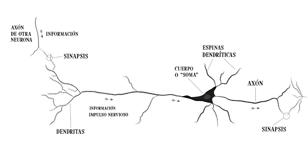
Diagram showing the parts of a neuron

The human brain has 86 billion neurons—as many as there are stars in the Milky Way. They communicate through electrical impulses and neurotransmitters. There are different types of neurons with different functions, but they all generally have a basic structure that includes: dendrites, which act as antennas to “listen” to other neurons—and sometimes other cells as well; the soma or cell body, which integrates the received information to transmit it to other cells and issue commands on what to do; and finally, the axon, which is a cable through which these messages travel to other cells at a speed of 360 km/h!

Diagram illustrating how a synapse works

SYNAPSE

Neurons communicate via electrical impulses through the movement of molecules and ions. When a neuron receives information from its dendrites, it generates an action potential that travels from the cell body to the tip of the axon. Neurons do not form a continuous network; rather, they are individual cells separated from one another by a small gap called the synaptic cleft. When it reaches the synapse, the nerve impulse must cross this gap to reach the cell that receives the information. When the nerve impulse reaches the end of the axon, the neuron releases neurotransmitters from its presynaptic terminal, and these reach the postsynaptic region—the part where the other cell receives the neurotransmitters and responds to them.

Neurotransmitters are molecules that neurons use to communicate. There are different neurotransmitters with very different functions, such as dopamine, which is associated with pleasure, and serotonin, which plays an important role in mood.

The existence of synapses could not be demonstrated until the development of the electron microscope in the mid-20th century, as can be seen in the image below.

Electron micrograph of a typical synapse (chemical synapse) between a parallel fibre and a dendritic spine of a Purkinje cell in the molecular layer of a mouse cerebellum.

CAJAL'S DRAWINGS

The garden of neurology offers researchers captivating insights and unparalleled artistic inspiration.

There, at last, my aesthetic instincts found complete satisfaction. Like an entomologist hunting for butterflies of striking hues, my attention sought out, in the orchard of grey matter, cells of delicate and elegant forms—the mysterious butterflies of the soul, whose fluttering wings might one day, who knows, shed light on the secret of mental life!

Cajal found in drawing the perfect tool for analysing, simplifying and schematising the stained specimens he observed through the microscope:

“There’s no doubt about it: only artists are drawn to science… I realised that to make a name for oneself with a paintbrush, one must turn one’s hand into a precision instrument. I owe who I am today to my artistic pursuits as a child—which my father strongly opposed. To date, I must have drawn more than 12,000 pictures. To the untrained eye, these are strange drawings, whose details are measured in thousandths of a millimetre, yet they reveal mysterious worlds within the architecture of the brain”.

Self-portrait at the microscope, c. 1910–1912

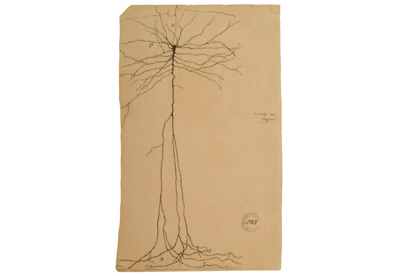
Santiago Ramón y Cajal, Giant deep pyramid of the ascending frontal circumvolution of Man, India ink on paper. Cajal-CSIC Legacy

BIOGRAFÍA