BIOGRAFÍA DE SANTIAGO RAMÓN Y CAJAL

CHAPTER 5

1884 1888

Neural theory

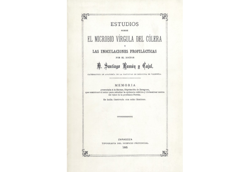
After being appointed to the chair of Anatomy at the University of Valencia, the family moved to the city in 1884, and Cajal began publishing Manual de histología normal y de técnica micrográfica (Manual of normal histology and micrographic technique) in instalments. The following year, he was commissioned by the Provincial Council of Zaragoza to conduct a study on cholera, which he carried out successfully. In 1887, he moved to Barcelona to take up a professorship in Normal and Pathological Histology. While serving as an examiner for competitive examinations in Madrid, he learnt the Golgi staining technique for the nervous system from Luis Simarro.

Zeiss Jena IVa Stativ Microscope, late 19th century. A. J. Schuhmacher Collection

Estudios sobre el microbio vírgula del cólera y las inoculaciones profilácticas (Studies on the cholera vibrio and prophylactic inoculations), Zaragoza, Provincial Hospice Printing Office, 1885

In 1885, commissioned by the Provincial Council of Zaragoza to study cholera, he conducted research and made discoveries regarding cholera vaccination at the Torre de los Canales in San Juan de Mozarrifar (Zaragoza). He developed a cholera vaccine by inoculating heat-killed germs. Cajal was the first person in the history of medicine to introduce the concept of a chemical vaccine that did not use live germs. However, this type of vaccine is credited to Salmon and Smith, who discovered it a year later. In recognition of his work, the Provincial Council of Zaragoza presented him with a high-quality Zeiss Stativ microscope, which proved instrumental in his subsequent research. “When I received that unexpected gift, I was overwhelmed with satisfaction and joy. Next to such a splendid Statif, with its abundance of objectives—including, among others, the famous 1.18 homogeneous immersion objective, which was then the state of the art in magnifying optics—my poor Verick microscope looked like a rickety old lock”. This microscope came to be known at the Cajal School as the “Zaragozano” (the Zaragoza model).

1888: “A CROWNING YEAR, A YEAR OF FORTUNE”

Building on Golgi’s method, Cajal proposed the neuron theory. “My crowning year, a year of good fortune, because during this year, which rises in my memory with the glow of dawn, those eagerly awaited and coveted discoveries finally emerged”.

Cajal describes the central nervous system as being composed of individual cells—neurons—that connect with one another but function as independent units, contrary to the accepted belief that it was a continuous network. Once his neural theory was firmly established, he turned his attention to the mechanism by which nerve impulses or currents travel. He focused his research on the retina and the olfactory bulb and developed his theory of the dynamic polarisation of nerve impulses.

The nerve impulse travels along the dendrites to the cell body and exits through the axon, which in turn connects to the dendrites of other nerve cells. Every neuron therefore has a receiving structure—the cell body or soma and the dendrites—a transmitting structure—the axon—and a distribution structure, which is the terminal nerve tree.

Self-portrait of Santiago Ramón y Cajal with a microtome; his Verick microscope can be seen in the background, c. 1884. Cajal-CSIC Legacy

HISTOLOGICAL TECHNIQUE

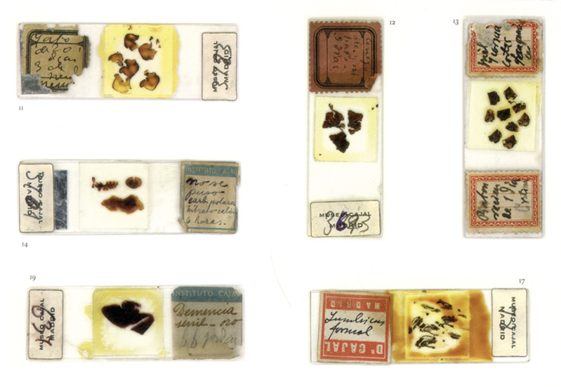
Histological techniques involve a series of laboratory procedures for preparing and studying biological tissues. This process includes fixing the tissue with chemical agents, dehydrating it, embedding it in paraffin or resin, cutting thin sections using a microtome, staining to highlight cellular structures and, finally, mounting the sections and examining them under a microscope.

From Luis Simarro, Cajal learnt the Golgi staining method of the time—the reazione nera—which consisted of fixing tissue fragments in a potassium dichromate solution for months. After that, following treatment with a silver nitrate solution, a small percentage of the cells were stained. Drawing on his knowledge of chemistry and his experiments with chemicals in the field of photography, Cajal added 2% osmium tetroxide to the potassium dichromate solution, accelerating the fixing and reaction process and reducing a procedure that used to take months to just one week.

He began his research by focusing on the cerebellum. Specifically, he studied young animals and embryos, whose nervous systems were still developing. He sensed that this would allow him to establish connections between the different regions of the brain and study how they develop in a simpler way.

Histological preparations made by Santiago Ramón y Cajal

The first illustration published by Cajal of a histological specimen (chicken cerebellum) stained using the Golgi method, 1888. Cajal-CSIC Legacy

An enlarged example of Golgi staining viewed under an electron microscope. Cajal-CSIC Legacy

BIOGRAFÍA